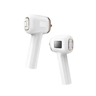
Fundus Camera
59,000 ฿
กล้องถ่ายภาพจอประสาทตาดิจิตอลแบบพกพา (Fundus Camera) มอบภาพถ่ายดวงตาสัตว์ที่คมชัด เพื่อการวินิจฉัยและเก็บประวัติระดับมืออาชีพ
• ถ่ายภาพจอประสาทตา (Fundus) ขั้วประสาทตา และเส้นเลือด ได้อย่างชัดเจนและมีสีสันสมจริง
• เทคโนโลยี Non-Mydriatic สามารถถ่ายภาพได้โดยไม่ต้องหยอดยาขยายรูม่านตา
• มีระบบลดเงาสะท้อนจากดวงตาสัตว์ (Tapetum lucidum) โดยเฉพาะ
• ภาพถ่ายสามารถนำมาอธิบายรอยโรคให้เจ้าของสัตว์เข้าใจได้ง่ายขึ้น
• บันทึกไฟล์ดิจิตอลลงคอมพิวเตอร์ผ่าน Wi-Fi หรือ USB เพื่อใช้ติดตามผลการรักษาในระยะยาว
คำอธิบาย
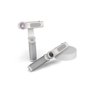
Fundus Camera สำหรับสัตวแพทย์ คือกล้องถ่ายภาพจอประสาทตาดิจิตอลแบบพกพา ที่ถูกสร้างขึ้นเพื่อเปลี่ยนการตรวจตาที่ซับซ้อนให้กลายเป็นการเก็บหลักฐานภาพถ่ายที่มีความละเอียดสูง เครื่องมือนี้ช่วยให้สัตวแพทย์สามารถจับภาพจอประสาทตา ขั้วประสาทตา และเส้นเลือด ได้อย่างรวดเร็วโดยไม่ต้องหยอดยาขยายรูม่านตา (Non-Mydriatic) ภาพถ่ายดิจิตอลที่ได้สามารถนำไปแสดงให้เจ้าของสัตว์ดูเพื่อความเข้าใจที่ตรงกัน รวมถึงสามารถบันทึกไว้เป็นประวัติการรักษาเพื่อติดตามการเปลี่ยนแปลงของโรคต้อหินหรือโรคจอประสาทตาเสื่อมในระยะยาวได้อย่างมีประสิทธิภาพ
ข้อมูลเชิงเทคนิคและคุณสมบัติเด่น:
ภาพถ่ายดิจิตอลความละเอียดสูง (High-Resolution Retinal Imaging): กล้องและเซนเซอร์ถูกออกแบบมาเป็นพิเศษเพื่อลดแสงสะท้อนจากชั้น Tapetum lucidum ในตาสัตว์ ให้ภาพจอประสาทตาที่คมชัดและสีสันสมจริง
เทคโนโลยีสแกนรูม่านตาขนาดเล็ก (Non-Mydriatic Technology): สามารถจับภาพผ่านรูม่านตาที่มีขนาดเล็กได้ ช่วยประหยัดเวลาในคลินิกและลดผลข้างเคียงจากการใช้ยาขยายรูม่านตา
หน้าจอและระบบจัดการข้อมูลอัจฉริยะ (Smart Display & Archiving): มีหน้าจอสีในตัวสำหรับเล็งและดูภาพทันที พร้อมระบบส่งต่อข้อมูลผ่าน Wi-Fi หรือ USB เพื่อเก็บลงในแฟ้มประวัติสัตว์ป่วย หรือส่งปรึกษาจักษุแพทย์เฉพาะทาง
The Portable Veterinary Fundus Camera is an advanced digital imaging system designed to transform complex ocular exams into highly documented, high-resolution evidence. This device empowers veterinarians to quickly capture stunning images of the retina, optic disc, and vasculature without the need for pupil-dilating drops (Non-Mydriatic). These crisp digital images can be instantly shared with pet owners for better clinical understanding and permanently archived in the patient’s medical record to effectively track the long-term progression of conditions like glaucoma or progressive retinal atrophy.
Key Technical Features & Specifications:
High-Resolution Retinal Imaging: The optics and digital sensors are specially calibrated to mitigate the intense reflections from the animal’s tapetum lucidum, producing incredibly sharp, true-color images of the fundus.
Non-Mydriatic Technology: Engineered to successfully capture wide-angle images through remarkably small, undilated pupils, saving valuable clinic time and eliminating the adverse effects of mydriatic eye drops.
Smart Display & Archiving: Features an integrated color screen for real-time targeting and image review. The device seamlessly transfers images via Wi-Fi or USB directly to the clinic’s patient management software or to off-site board-certified ophthalmologists for consultation.