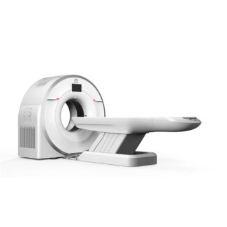
CBCT TC-460
5,990,000 ฿
เครื่องเอกซเรย์คอมพิวเตอร์แบบ Cone Beam รุ่น CBCT TC-460 มอบภาพวินิจฉัย 3 มิติที่มีความละเอียดสูงสุด ออกแบบมาเฉพาะสำหรับงานทันตกรรมสัตว์ ศัลยกรรมบริเวณใบหน้า และการรักษาสัตว์แปลก (Exotic pets)
• เทคโนโลยี Cone Beam 3D สร้างภาพโครงสร้างเนื้อเยื่อแข็ง กระดูก และรากฟันได้อย่างแม่นยำทุกมิติ
• ให้ภาพความละเอียดสูง (Isotropic resolution) โดยใช้ปริมาณรังสีที่ต่ำกว่า CT Scan ทั่วไปอย่างมาก
• ดีไซน์กะทัดรัด ประหยัดพื้นที่ ติดตั้งได้ง่ายในคลินิกทันตกรรมสัตว์หรือโรงพยาบาลสัตว์ที่มีพื้นที่จำกัด
• ตอบโจทย์การวินิจฉัยโรคปริทันต์ที่ซับซ้อน ฝีที่รากฟัน กระดูกขากรรไกรหัก และความผิดปกติในโพรงจมูก
• มาพร้อมซอฟต์แวร์ประมวลผล 3 มิติเฉพาะทาง และรองรับมาตรฐาน DICOM 3.0 อย่างสมบูรณ์แบบ
คำอธิบาย
CBCT TC-460 (Cone Beam Computed Tomography) คือเครื่องเอกซเรย์คอมพิวเตอร์แบบกรวยรังสีที่เข้ามาพลิกโฉมวงการทันตกรรมและศัลยกรรมสรีระใบหน้าของสัตว์เลี้ยง ในขณะที่การเอกซเรย์ฟันแบบ 2 มิติทั่วไปมักมีข้อจำกัดเรื่องภาพซ้อนทับกัน TC-460 สามารถเก็บข้อมูลภาพ 3 มิติของกะโหลกศีรษะ ขากรรไกร หรือขาของสัตว์ได้ครบถ้วนในการหมุนเพียงรอบเดียว ช่วยให้สัตวแพทย์และทันตแพทย์สัตว์สามารถมองเห็นความลึก โครงสร้างรากฟัน กระดูกเบ้าฟัน และแนวเส้นประสาทได้อย่างชัดเจน นำไปสู่การวางแผนถอนฟันที่ยากลำบากหรือการผ่าตัดใบหน้าได้อย่างปลอดภัยและแม่นยำขั้นสุด
ข้อมูลเชิงเทคนิคและคุณสมบัติเด่น:
เทคโนโลยีกรวยรังสีและรังสีต่ำ (Advanced Cone Beam Technology): แตกต่างจากเครื่อง CT Scan ทั่วไปที่ใช้ลำแสงแบบพัด (Fan-beam) เครื่อง TC-460 จะยิงลำแสงเอกซเรย์เป็นรูปกรวย (Cone-shaped) เพื่อเก็บภาพพื้นที่ที่ต้องการทั้งหมดในการหมุนรอบเดียว วิธีนี้ไม่เพียงแต่ช่วยให้สแกนเสร็จรวดเร็ว (ลดเวลาการวางยาสลบสัตว์) แต่ยังช่วยลดปริมาณรังสีที่สัตว์และผู้ปฏิบัติงานจะได้รับลงอย่างมหาศาล
ความคมชัดสูงสุดสำหรับงานทันตกรรม (Unmatched Dental & Maxillofacial Clarity): ระบบถูกออกแบบมาให้โดดเด่นในการถ่ายภาพเนื้อเยื่อแข็ง โดยให้ความละเอียดในระดับซับมิลลิเมตร (Sub-millimeter) ซึ่งความละเอียดระดับนี้มีความสำคัญอย่างยิ่งต่อการค้นหารอยโรคที่มักถูกมองข้ามในการเอกซเรย์ปกติ เช่น โรคฟันผุกร่อนในแมว (FORLs), รอยร้าวขนาดเล็กที่ข้อต่อขากรรไกร (TMJ), และการติดเชื้อที่ปลายรากฟันลึกๆ
ดีไซน์กะทัดรัด ติดตั้งง่าย (Compact & Clinic-Friendly Design): ข้อได้เปรียบที่สำคัญที่สุดอย่างหนึ่งของ CBCT TC-460 คือขนาดที่กะทัดรัด ตัวเครื่องใช้พื้นที่ในการติดตั้งและต้องการการกรุผนังกันรังสีน้อยกว่าเครื่อง CT Scan แบบ 64 สไลซ์ทั่วไปมาก ทำให้คลินิกรักษาสัตว์ขนาดกลางหรือศูนย์ทันตกรรมเฉพาะทางสามารถนำเทคโนโลยี 3 มิติระดับท็อปมาใช้ในคลินิกได้โดยไม่ต้องปรับปรุงพื้นที่ครั้งใหญ่
ซอฟต์แวร์ประมวลผล 3 มิติและการเชื่อมต่อ (Comprehensive 3D Software & DICOM Integration): มาพร้อมซอฟต์แวร์เวิร์กสเตชันที่ทรงพลัง สามารถสร้างภาพตัดขวางหลายระนาบ (MPR) และภาพโมเดล 3 มิติได้ในเวลาไม่กี่วินาที สัตวแพทย์สามารถหมุนดูและผ่าโมเดล 3 มิติได้จากทุกแกน (Axial, Coronal, Sagittal) เพื่อวัดขนาดได้อย่างแม่นยำ นอกจากนี้ตัวเครื่องยังรองรับมาตรฐาน DICOM ทำให้สามารถส่งต่อข้อมูล 3 มิติขนาดใหญ่ไปยังเซิร์ฟเวอร์ PACS หรือส่งให้สัตวแพทย์เฉพาะทางภายนอกวิเคราะห์ได้อย่างไร้รอยต่อ
The CBCT TC-460 (Cone Beam Computed Tomography) is a game-changing diagnostic tool that elevates veterinary dentistry and localized orthopedic imaging to an entirely new level. While traditional 2D dental X-rays are limited by overlapping structures, the TC-460 captures a complete, distortion-free 3D volume of the animal’s skull, jaw, or limbs in a single rotation. This allows veterinary dentists and surgeons to examine the exact spatial relationship of tooth roots, the alveolar bone, and critical neurovascular canals, ensuring absolute precision when planning complicated extractions or maxillofacial surgeries.
Key Technical Features & Specifications:
Advanced Cone Beam Technology: Unlike conventional fan-beam CT scanners that capture data slice by slice, the TC-460 utilizes a cone-shaped X-ray beam to capture the entire target region in one quick rotation. This highly efficient acquisition method not only speeds up the scanning process—reducing the time the animal is under anesthesia—but also drastically lowers the overall radiation exposure to the patient and staff.
Unmatched Dental & Maxillofacial Clarity: Designed specifically to excel in high-contrast hard tissue imaging, the system provides sub-millimeter isotropic resolution. This extreme level of detail is critical for detecting subtle pathologies that are often missed on standard radiographs, such as Feline Odontoclastic Resorptive Lesions (FORLs), micro-fractures in the temporomandibular joint (TMJ), and deep-seated root tip infections.
Compact & Clinic-Friendly Design: One of the greatest advantages of the CBCT TC-460 is its minimal footprint. It requires significantly less room and specialized lead shielding compared to a standard 64-slice CT scanner, allowing average-sized clinics and specialized veterinary dental centers to offer premium 3D imaging in-house without the need for extensive facility renovations.
Comprehensive 3D Software & DICOM Integration: The system comes paired with powerful, intuitive workstation software capable of generating Multi-Planar Reconstructions (MPR) and 3D Volume Rendering in seconds. Veterinarians can slice through the 3D model in any plane (axial, coronal, sagittal) for precise measurements. Fully compliant with DICOM standards, the 3D data can be effortlessly shared with teleradiology specialists or integrated into the hospital’s PACS network.